The Australian Government is now in caretaker period. During this time, updates on this website will be published in accordance with the Guidance on Caretaker Conventions, until after the election.
NHMRC's Science to Art Award recognises outstanding imagery that has arisen from research funded by the NHMRC.
Imaging is now an integral technology in medical research. These images are not only scientifically important, but they can also be aesthetically powerful. The winner is selected by the Council of NHMRC and receives a framed print of their image and acknowledgement of the achievement by NHMRC.
Entries
Entries for the 2027 Science to Art Award will open in 2026. For queries about this award, contact nhmrc.awards@nhmrc.gov.au.
Winners
2025 - Associate Professor Arutha Kulasinghe

Description
This is a digital image of skin cancer located in the head and neck region. The skin cells are shown in bright red, with the tumour pockets in dull red surrounded by yellow. Immune cells (including macrophages and lymphocytes) are patrolling the tissue in green and magenta.
The image was captured using spatial proteomics, which was the 2024 Nature Method of the Year, and allows the visualisation of up to 100 biomarkers at the single cellular level across the entire tissue section. Referred to as the ‘Google Maps’ approach to tissue profiling, this revolutionary approach can be used to map every cell across different tumour types and tissues to unravel why some tumours respond to treatment while others do not.
Better understanding of the tumour microenvironment for each individual patient would allow for treatment and therapy personalisation to improve the care and outcomes for individuals affected by cancer.
2023 – Dr Caleb Dawson

Description
This is an image of just 1 mm of healthy breast tissue in lactation. The tiny yellow (F-actin) and magenta (magenta) muscle-like myoepithelial cells are wrapping around milk-producing spheres, called alveoli. The blood vessel running vertically delivers oxytocin that is released upon breastfeeding. Oxytocin causes the muscle-like cells to contract and squeeze out the milk. This image was captured using a confocal microscope after staining and clearing the tissue. Images like this have helped discover a unique breast immune cell that removes the milk-producing cells after weaning to keep the breast healthy.
2021 – Professor Frederic Hollande

Description
This image depicts a population of metastatic colorectal cancer cells which have been 'optically barcoded' with different fluorescence markers. By tracking the different coloured tumour cells over time, the research team can monitor how individual cells respond to chemotherapy and use this knowledge to study mechanisms of drug resistance in cancer.
2019 – Mr Jianqun Gao

Description
Human neurons differentiated from neural stem cells (NSCs) have been used as a novel disease model to study neurodegenerative diseases. This image is showing nuclei (blue) of these NSC-derived neurons, as well as their networks (green and red). It looks like an eddy of neurons attracting us to discover an unknown world. The experiments conducted with these neurons hope to unravel the mechanisms of neurodegenerative diseases, such as Parkinson's disease.
2017 – Joshua SS Li
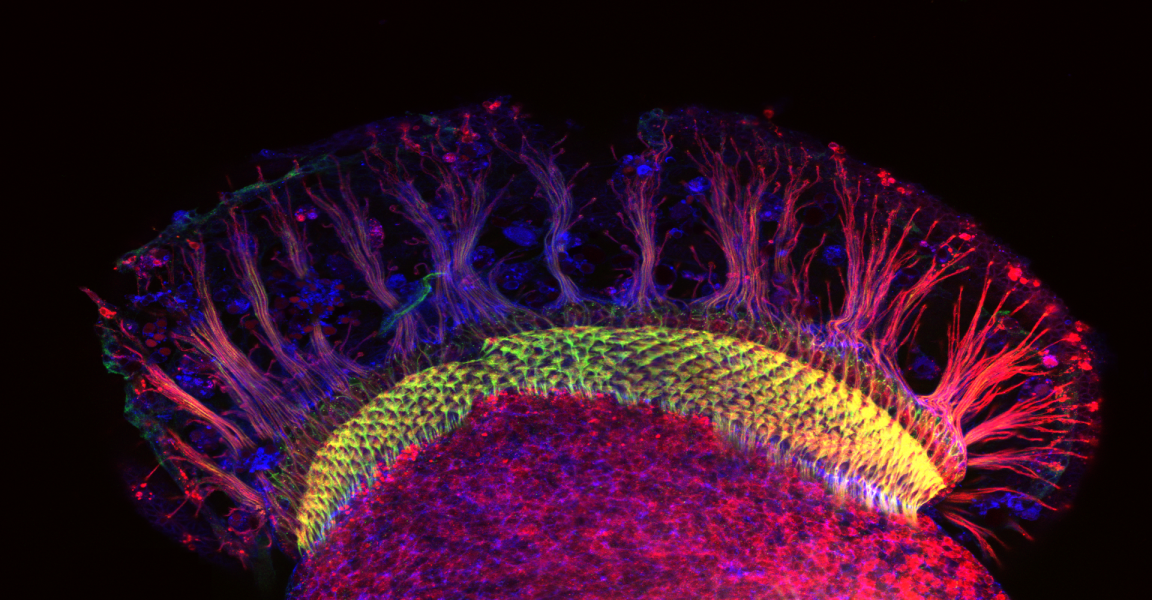
Description
In the developing brain, neurons find their prospective partners with high precision. How this complex form of biological hardwiring is achieved remains elusive. This image is of the developing visual system of a fruit fly dissected, fixed, immunostained and whole-mounted. Photoreceptor neurons (top) project down towards the lamina neuropil (green) to establish synapses with specific partners. Experiments conducted in this system hope to unravel mechanisms of neurodevelopment.
2015 – Victor Anggono

Description
Developing nerve cells (neurons) extend their processes, known as axons, and project to one another to establish synaptic connection and form a neural circuit, the activity of which is essential for brain function such as learning and memory.
2013 – Dr Michael Lovelace and colleagues

Description
Laser therapy is currently used to treat chronic pain in patients worldwide. This study aimed to elucidate the cellular mechanisms involved in the response of neurons to laser irradiation. This image depicts a monolayer of cultured dorsal root ganglion neurons (DRGs), Schwann and satellite cells, used in modelling the response of neurons to laser irradiation. Dye labeling of the cell membranes allows fine processes extending between DRGs to be visualized, while separately we investigated changes in mitochondrial membrane potential. Collectively, these experiments have allowed us to broaden our understanding of the cellular mechanisms involved in pain relief through laser therapy.
2011 – Eric Hanssen
Description
These structures are responsible for the export of the principal virulence factor of the parasite. This segmentation model was made with IMOD from an electron tomogram of a whole Plasmodium falciparum parasite. The rendering was done with Blender.