
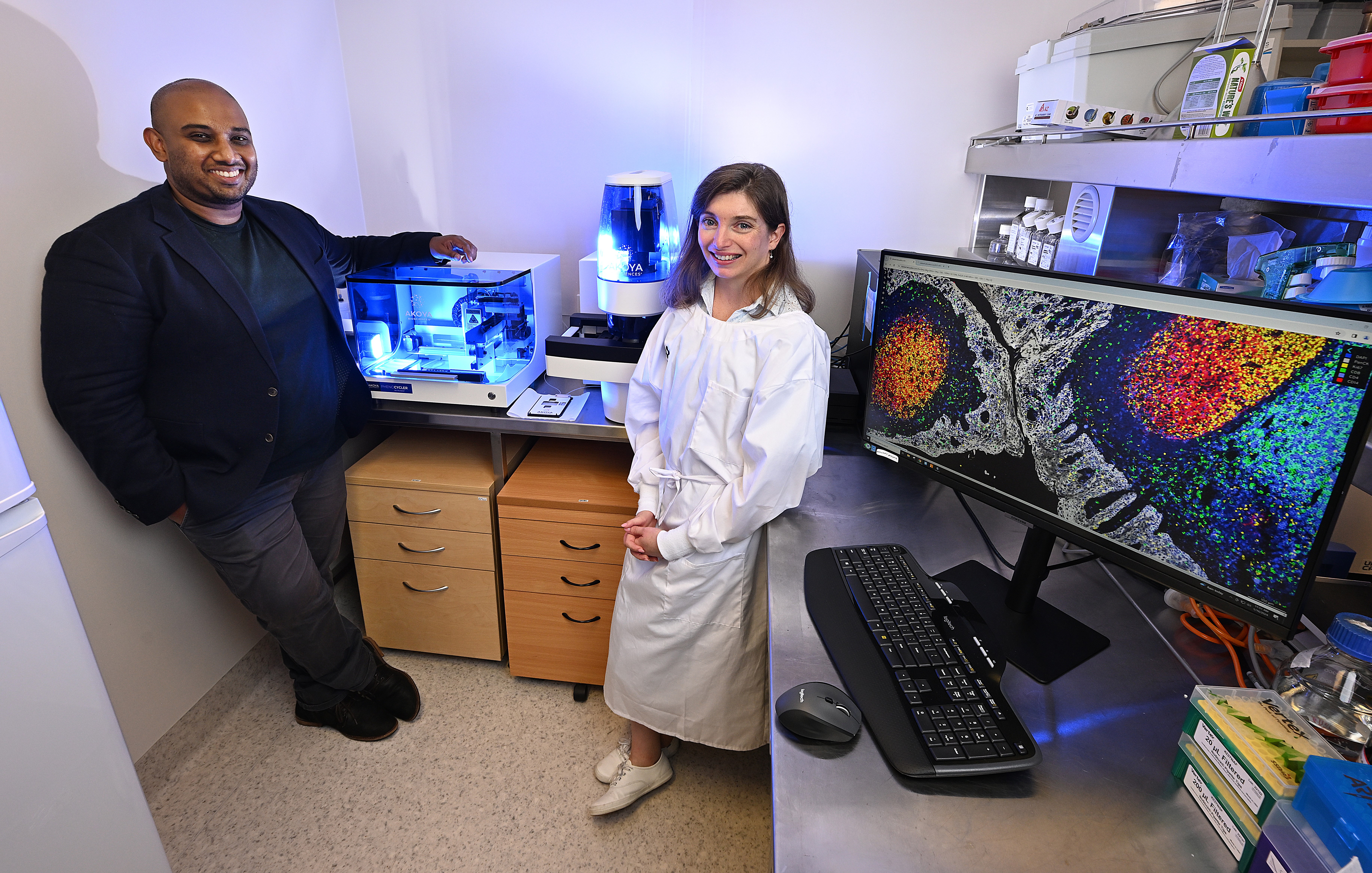
Associate Professor Arutha Kulasinghe, recipient of the 2025 NHMRC Science to Art Award, is bringing together the rigour of heath and medical research with aesthetically powerful art. With a focus on cutting edge spatial biology and genomic techniques, Associate Professor Kulasinghe is dedicated to unlocking the secrets of individual cell interactions, leading to personalised treatments for several diseases, including cancer.
Associate Professor Kulasinghe leads the Clinical-oMx Lab at the Frazer Institute, University of Queensland and is the Founding Scientific Director of the Queensland Spatial Biology Centre. Associate Professor Kulasinghe's innovative research aims to map each patient's tumour at the cellular level for lung, skin, and head and neck cancers using next generation digital profiling techniques.

‘Battleground beneath the skin’ the winning artwork for the 2025 NHMRC Science to Art Award represents Associate Professor Kulasinghe’s and his team’s efforts in understanding the tumour biology of each patient, in a cell-by-cell manner, to better create digital maps for interrogation for therapeutic efficacy. With a focus on lung cancer, head and neck cancer and skin cancer, this technique is helping to define why next generation immunotherapies are effective in only some patients, to ultimately tailor therapies for those likely to respond.
When asked what he hopes this research will achieve, Associate Professor Kulasinghe hopes it will lead to “the development of advanced diagnostics and therapeutics for cancer patients where the therapies that are given are based on the individual patient’s tumour profile, enabling a personalised medicine approach.”
“Cancer patients, diagnosed with lung cancer, head and neck or skin cancer, will gain the most benefits from this research, but the methodology and analysis are applicable to melanoma, bladder and breast cancers (most solid cancers),” says Associate Professor Kulasinghe.
Reflecting on why he chose this area of research, Associate Professor Kulasinghe said that his involvement in the care of his grandfather (who had advanced stage colorectal cancer) increased his understanding of needing better diagnostics and therapeutics for cancer patients. This helped him define his ‘why’.
When asked how this research contributes to building a healthy Australia, and a healthy globe, Associate Professor Kulasinghe said that it provides a greater depth of understanding of each patient’s tumour for Australia’s most lethal cancers.
“This type of tumour profiling is world leading and likely to aid in the development of better diagnostics and therapeutics for Australians and global citizens,” says Associate Professor Kulasinghe.
Associate Professor Kulasinghe has pioneered spatial transcriptomics, proteomics and interactomics in the Asia Pacific region with funding support from the MRFF, NHMRC, US DoD, Cancer Australia, Cure Cancer and numerous hospital and philanthropic organisations. He is also the recipient of Cure Cancer’s 2023 Researcher of the Year Award and was included in The Australian’s Top Innovators List in 2024.
Pertinent to receiving NHMRC’s 2025 Science to Art Award, Associate Professor Kulasinghe’s cancer imagery has been wrapped in custom Xboxes and auctioned to raise money for cancer research with a global reach of over 170 million people.
To Associate Professor Kulasinghe, the key driver of this level of research success for him is team science.
“We no longer live in silos, our research needs the compliment of technologists, immunologists, pathologists, medical oncologists, computational biologists, data scientists and patient advocates,” he said.
Representative of this is Battleground beneath the skin, a digital image of skin cancer located in the head and neck region. The image was captured using spatial proteomics, the 2024 Nature Method of the Year, and allows the visualisation of up to 100 biomarkers at the single cellular level across the entire tissue section.
Referred to as the ‘Google Maps’ approach to tissue profiling, this revolutionary approach can be used to map every cell across different tumour types and tissues to unravel why some tumours respond to treatment while others do not. Better understanding of the tumour microenvironment for each individual patient would allow for treatment and therapy personalisation to improve the care and outcomes for individuals affected by cancer.
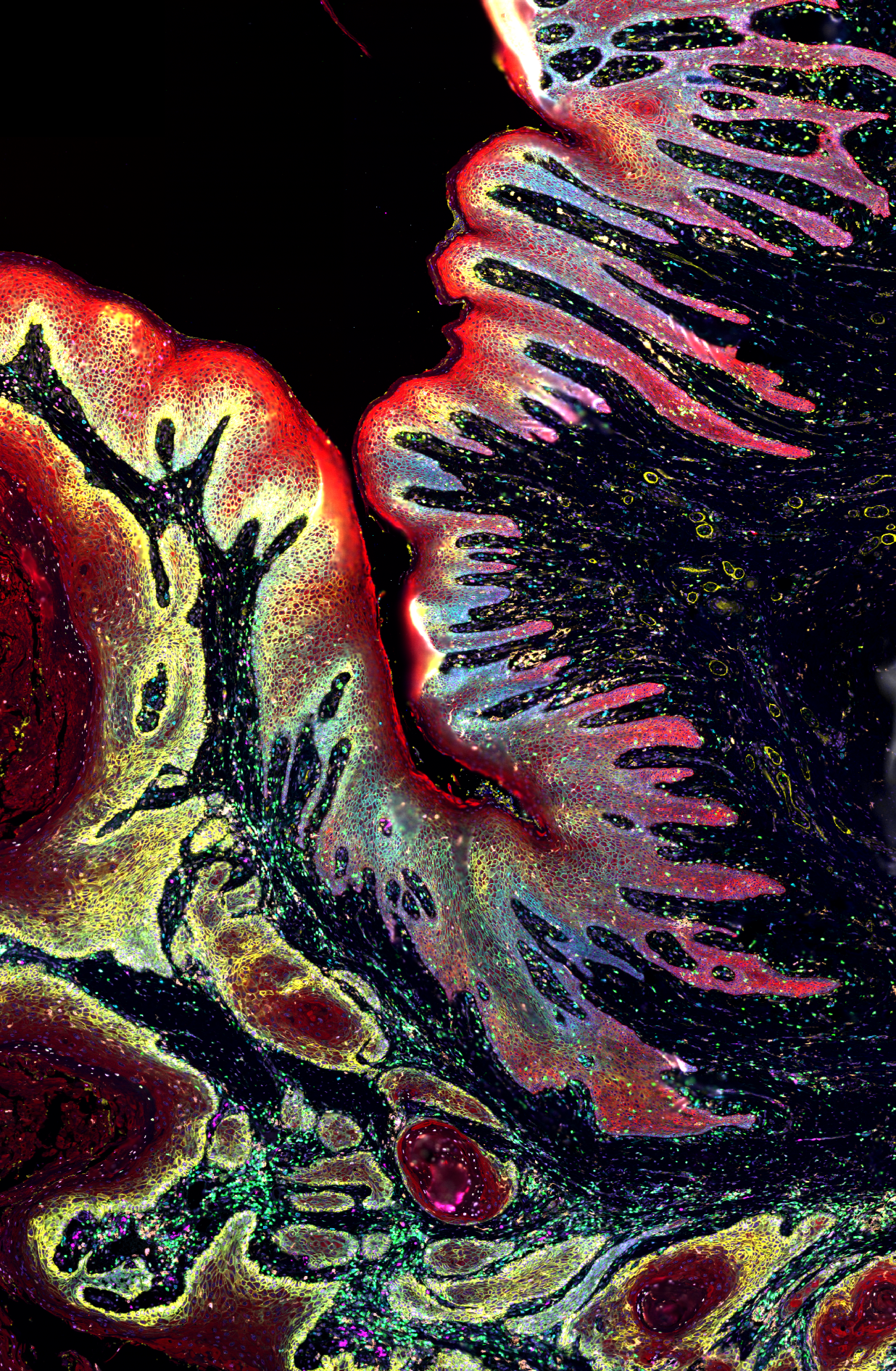
Battleground beneath the skin from Associate Professor Arutha Kulasinghe, Frazer Institute, University of Queensland.
The skin cells are shown in bright red, with the tumour pockets in dull red surrounded by yellow. Immune cells (including macrophages and lymphocytes) are patrolling the tissue in green and magenta.